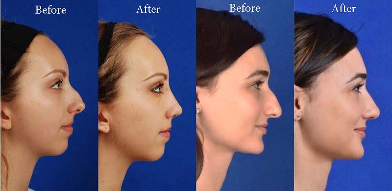
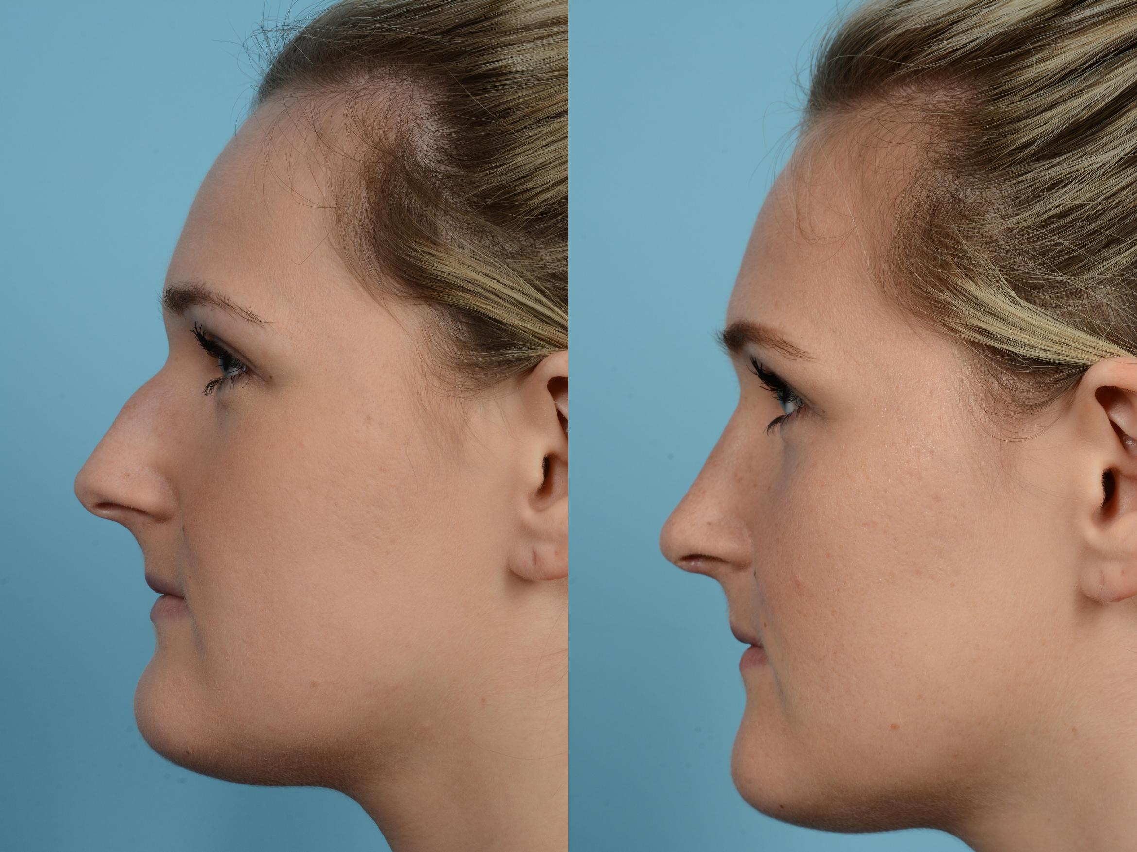
How Rhinoplasty Austin Tx can Save You Time, Stress, and Money.
Table of ContentsNot known Incorrect Statements About Austin Rhinopasty Surgeon Rhinoplasty Surgeon Austin Fundamentals ExplainedExcitement About Rhinoplasty Surgeon Austin
Based upon the available area of nasal skin, the surgeon picks the place for the bilobed flap, and orients the pedicle. If the flaw is in the lateral element of the nose, the pedicle is based medially. If the defect is at the nasal idea, or at the nasal dorsum, the pedicle is based laterally.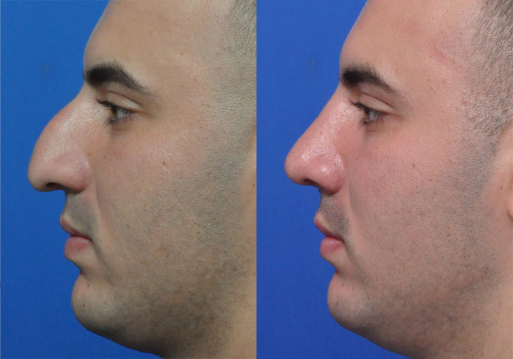
The outer semi-circle defines the needed length of the two lobes of the skin flap. The inner semi-circle bisects the center of the initial wound, and continues throughout the donor skin, establishing limit procedure of the pedicle typical to the two lobes of the flap. The cosmetic surgeon then draws two lines from the peak of the injury; the first line drawn is at an angle of 45 degrees from the long axis of the injury, and the second line drawn is at a 90-degree angle from the axis of the injury.
The delineation of each of the 2 lobes of the flap begins and ends at the inner semi-circle, and encompasses the outer semi-circle, to the point where it intersects its main axis. The width of the first lobe is around 2 mm narrower than the width of the wound; the width of the 2nd lobe is roughly 2 mm narrower than the width of the first lobe.
The injury is deepened, to the nasal skeleton, to accommodate the tissue density of the bilobed flap (rhinoplasty surgeon austin). Technically, cutting the injury, enlarging it, is preferable, and much safer, than cutting (thinning) the flap to fit the injury. Weakening the donor site for the 2nd lobe allows closing it mainly; it likewise removes excess-skin "dog-ears" at the donor site.
II. Nasolabial flap In the 19th century, the surgical methods of J.F. Dieffenbach (17921847) popularized the nasolabial flap for nasal restoration, for which it stays a fundamental nose surgery treatment. The nasolabial flap can be either superiorly based or inferiorly based; of which the superiorly based flap is the more practical rhinoplastic application, because it has a more versatile arc of rotation, and the donor-site scar is inconspicuous.
Some Known Details About Rhinoplasty Austin Tx
The blood supply for the flap pedicle are the transverse branches of the contralateral angular artery (the facial artery terminus parallel to the nose), and by a confluence of capillary from the angular artery and from the supraorbital artery in the medial canthus, (the angles formed by the conference of the upper and lower eyelids) (austin rhinopasty surgeon).
The nasolabial flap is a random flap that is emplaced with the proximal (near) portion resting upon the lateral wall of the nose, and the distal (far) portion resting upon the cheek, which contains the primary angular artery, and so is perfused with retrograde arterial circulation. Surgical method the nasolabial flap The pedicle of the nasolabial flap rests upon the lateral nasal wall, and is transposed an optimum of find out 60 degrees, in order to avoid the "bridge impact" of a flap emplaced across the nasofacial angle.
The shape of the skin flap is cut from the wound template produced by the cosmetic surgeon. An incision is made to the flap (without an anaesthetic injection of epinephrine), which then is elevated and oriented, in an inferior-to-superior instructions, in between the subcutaneous fat and the muscle fascia. The cutting continues up until the skin flap can be freely transposed upon the try this site nasal problem.
The flap then is bent back (reflected), and can be thinned (cut) under loupe magnification; nevertheless, a nasolabial flap can not be thinned as quickly as an axial skin-flap. rhinoplasty austin tx. After the nasolabial flap has actually been emplaced, the flap donor-site wound is sutured closed. For an injury of the lateral nasal wall that is less than 15 mm broad, the flap donor-site can be closed mostly, with stitches.
Such dangers are avoided by advancing (moving) the skin of the cheek towards the nasofacial junction, where it is sutured to the deep tissues. Moreover, a narrow wound, less than 1 mm broad can be permitted to recover by secondary intention (autonomous re-epithelialisation). III. The paramedian forehead flap The paramedian forehead flap is the premier autologous skin graft for the restoration of a nose, by replacing any of the aesthetic nasal subunits, specifically concerning the issues of various tissue thickness and skin color.
Limited length is an useful application limit of the paramedian forehead flap, specifically when the client has a low frontal hairline. In such a patient, a little portion of scalp skin can be included to the flap, however it does have a various skin texture and does continue growing hair; such mismatching is avoided with the transverse emplacement of the flap along the hairline; yet that part of the skin flap is random, therefore runs the risk of a higher incidence of necrosis.
The smart Trick of Austin Rhinopasty Surgeon That Nobody is Talking About
Nevertheless, the get redirected here second stage of the nasal reconstruction can be carried out with the client under regional anaesthesia. Aesthetically, although the flap donor-site scar heals well, it is visible, and thus hard to conceal, specifically in men. Nose job: A paramedian forehead flap design. Surgical method the paramedian forehead flap The surgeon creates the paramedian forehead flap from a custom-fabricated three-dimensional metal foil template stemmed from the measures of the nasal problem to be remedied.
Later on, the distal half of the flap is dissected and thinned to the subdermal plexus. The surgeon makes a metal foil design template stemmed from the measurements of the nasal wound. Using a Doppler ultrasonic scanner, the cosmetic surgeon determines the axial pedicle of the tissue-flap (composed of the supraorbital artery and the supratrochlear artery), generally at the base, next to the median eyebrow; the point usually is in between the midline and the supraorbital notch.